Clinical videos + HTML notes for NEET PG-ready revision.
Streamora lets you upload case-based videos, attach rich HTML notes, like and comment on other clinicians’ posts, follow creators and save structured exam-oriented pearls – all mobile-first and SEO-ready.
Why Streamora stands out for NEET PG aspirants: Unlike generic apps, we focus on high-yield topics like labour stages (from first to fourth, with management pitfalls), Rh-negative pregnancies (anti-D prophylaxis timing), and anatomy pearls (e.g., sacral hiatus for caudal blocks). Our community-driven notes include real residency cases—e.g., "Missed Rh isoimmunization leading to hydrops fetalis"—with attached flowcharts and MCQ-style tags for quick revision. Free, ad-light, and built by med students for med students.
Latest Medical Posts
High-yield medical discussions, NEET PG-flavoured notes and short clinical cases from all users.
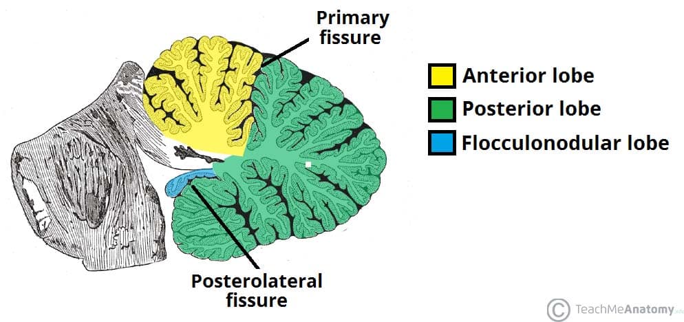
Cerebellum Anatomy Structure Functions and Clinical Importance
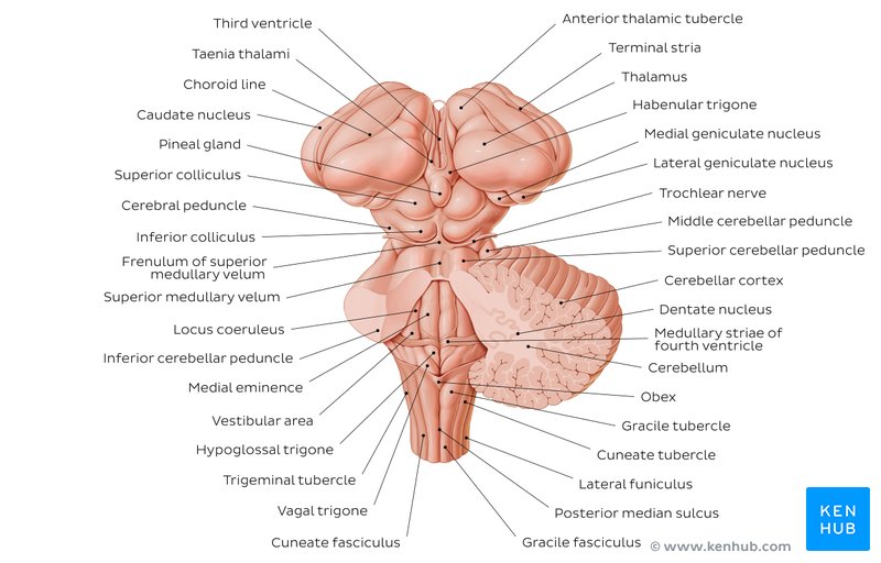
## Cerebellum Anatomy – Complete SEO-Friendly Guide ### Introduction The **cerebellum** is a major part of the hindbrain that plays a critical role in **coordination of movement, balance, posture, muscle tone, and motor learning**. Although it does not initiate movement, it fine-tunes motor activity to ensure accuracy and smooth execution. --- ## Location and Relations * Situated in the **posterior cranial fossa** * Lies **behind the pons and medulla** * Separated from the cerebrum by the **tentorium cerebelli** * Forms the **roof of the fourth ventricle** --- ## Gross Anatomy of the Cerebellum ### External Features The cerebellum consists of: 1. **Two hemispheres** (right and left) 2. **Vermis** (midline structure connecting hemispheres) #### Surfaces * **Superior surface** * **Inferior surface** * Both surfaces show numerous transverse folds called **folia** --- ## Lobes of the Cerebellum The cerebellum is divided by fissures into **three lobes**: ### 1. Anterior Lobe * Located anterior to the **primary fissure** * Functionally related to **spinocerebellum** * Involved in **posture and gait control** ### 2. Posterior Lobe * Largest lobe * Lies between primary fissure and posterolateral fissure * Involved in **fine voluntary movements** ### 3. Flocculonodular Lobe * Composed of **flocculus + nodulus** * Also called **vestibulocerebellum** * Responsible for **balance and eye movements** --- ## Functional Divisions of the Cerebellum ### 1. Cerebrocerebellum * Lateral hemispheres * Connected to cerebral cortex * Controls **planning and coordination of skilled movements** ### 2. Spinocerebellum * Vermis and intermediate zones * Regulates **muscle tone and ongoing movements** ### 3. Vestibulocerebellum * Flocculonodular lobe * Maintains **equilibrium and eye coordination** --- ## Cerebellar Cortex (Microscopic Anatomy) ### Layers of Cerebellar Cortex The cerebellar cortex has **three layers**: 1. **Molecular Layer** * Contains stellate and basket cells * Few neurons, mostly fibers 2. **Purkinje Cell Layer** * Single layer of large **Purkinje cells** * Output neurons of the cerebellar cortex * Inhibitory (GABAergic) 3. **Granular Layer** * Contains granule cells and Golgi cells * Highly cellular --- ## White Matter of Cerebellum * Located deep to the cortex * Appears as **arbor vitae** (tree-like pattern) * Carries fibers connecting cortex to cerebellar nuclei --- ## Deep Cerebellar Nuclei Embedded within white matter: 1. **Dentate** 2. **Emboliform** 3. **Globose** 4. **Fastigial** These nuclei serve as **major output centers** of the cerebellum. --- ## Cerebellar Peduncles The cerebellum connects to the brainstem via **three paired peduncles**: ### 1. Superior Cerebellar Peduncle * Connects to **midbrain** * Mainly **efferent (output)** fibers ### 2. Middle Cerebellar Peduncle * Connects to **pons** * Largest peduncle * Carries **afferent fibers from cerebral cortex** ### 3. Inferior Cerebellar Peduncle * Connects to **medulla** * Mixed afferent and efferent fibers --- ## Blood Supply of the Cerebellum * **Superior cerebellar artery (SCA)** * **Anterior inferior cerebellar artery (AICA)** * **Posterior inferior cerebellar artery (PICA)** All arise from the **vertebrobasilar system**. --- ## Functional Summary * Coordinates voluntary movements * Maintains posture and balance * Regulates muscle tone * Involved in motor learning and error correction --- ## Clinical Correlation (Brief) Lesions of the cerebellum lead to: * **Ataxia** * **Dysmetria** * **Intention tremor** * **Nystagmus** * **Hypotonia** * **Scanning speech** --- ## SEO Title **Cerebellum Anatomy Structure Functions and Clinical Importance** ## Meta Description Comprehensive SEO-friendly guide on cerebellum anatomy covering location lobes functional divisions cerebellar cortex peduncles blood supply and clinical significance. ## SEO Keywords cerebellum anatomy, cerebellar lobes, cerebellar cortex layers, cerebellar peduncles, deep cerebellar nuclei, cerebellum functions, hindbrain anatomy, neuroanatomy cerebellum --- If you want next: * **30 case-based cerebellar lesion scenarios** * **Hard MCQs with explanations** * **Clinical syndromes of cerebellar lesions** * **HTML/CSS/JS exam-mode MCQs** Just tell me 👍 Below are **high-yield clinical case scenarios of cerebellar lesions with clear diagnosis and management**, written in an **exam-oriented, clinical approach**. Concise but complete. --- ## 1. Midline (Vermis) Lesion – Truncal Ataxia **Case** A 45-year-old man with chronic alcohol use presents with inability to sit or stand without support. Limb movements are relatively normal. Gait is broad-based and staggering. **Clinical Diagnosis** Vermian cerebellar lesion (anterior lobe degeneration) **Key Features** * Truncal ataxia * Broad-based gait * Minimal limb ataxia **Management** * Treat underlying cause (alcohol cessation) * Thiamine supplementation * Physiotherapy for gait and balance * Fall-prevention measures --- ## 2. Cerebellar Hemisphere Lesion – Ipsilateral Limb Ataxia **Case** A 60-year-old man with hypertension presents with clumsiness of the right hand. Finger-nose test shows past pointing on the right. **Clinical Diagnosis** Right cerebellar hemisphere infarction **Key Features** * Ipsilateral limb ataxia * Dysmetria * Intention tremor **Management** * MRI brain to confirm stroke * Antiplatelet therapy * Blood pressure and risk factor control * Neurorehabilitation --- ## 3. Flocculonodular Lobe Lesion – Balance Disorder **Case** A child presents with frequent falls, vertigo, and abnormal eye movements. **Clinical Diagnosis** Vestibulocerebellar lesion **Key Features** * Nystagmus * Vertigo * Severe balance impairment **Management** * Treat underlying cause (tumor/infection) * Vestibular rehabilitation * Anti-vertigo medications (short term) --- ## 4. Acute Cerebellar Stroke **Case** A 70-year-old patient presents with sudden onset vertigo, vomiting, ataxia, and headache. **Clinical Diagnosis** Cerebellar infarction (PICA/AICA territory) **Management** * Emergency CT/MRI brain * Manage raised intracranial pressure * Antiplatelet or anticoagulation as indicated * Neurosurgical decompression if brainstem compression --- ## 5. Cerebellar Hemorrhage **Case** A hypertensive patient develops sudden headache, vomiting, and rapid deterioration of consciousness. **Clinical Diagnosis** Cerebellar hemorrhage **Management** * Immediate CT brain * Blood pressure control * Neurosurgical evacuation if large bleed * ICU monitoring --- ## 6. Alcoholic Cerebellar Degeneration **Case** A chronic alcoholic presents with progressive gait instability over months. **Clinical Diagnosis** Anterior cerebellar lobe degeneration **Management** * Alcohol abstinence * Nutritional rehabilitation * Thiamine and multivitamins * Long-term physiotherapy --- ## 7. Multiple Sclerosis with Cerebellar Involvement **Case** A young woman presents with intention tremor, scanning speech, and nystagmus. **Clinical Diagnosis** Cerebellar involvement in multiple sclerosis **Management** * MRI brain with contrast * Acute relapse: corticosteroids * Disease-modifying therapy * Speech and occupational therapy --- ## 8. Cerebellar Tumor (Medulloblastoma) **Case** A child presents with morning vomiting, headache, and gait ataxia. **Clinical Diagnosis** Midline cerebellar tumor (medulloblastoma) **Management** * MRI brain * Surgical excision * Radiotherapy and chemotherapy * Long-term neurodevelopmental follow-up --- ## 9. Cerebellar Abscess **Case** A patient with chronic otitis media presents with fever, headache, and ataxia. **Clinical Diagnosis** Cerebellar abscess **Management** * MRI with contrast * IV broad-spectrum antibiotics * Surgical drainage if indicated * Treat source of infection --- ## 10. Drug-Induced Cerebellar Toxicity **Case** A patient on phenytoin presents with nystagmus and ataxia. **Clinical Diagnosis** Drug-induced cerebellar dysfunction **Management** * Check drug levels * Stop or reduce offending drug * Supportive care * Monitor recovery --- ## 11. Paraneoplastic Cerebellar Degeneration **Case** A middle-aged woman presents with rapidly progressive ataxia; later found to have ovarian carcinoma. **Clinical Diagnosis** Paraneoplastic cerebellar degeneration **Management** * Treat underlying malignancy * Immunotherapy (steroids, IVIG) * Supportive rehabilitation --- ## 12. Cerebellar Ataxia in Hypothyroidism **Case** A patient presents with slow speech, unsteady gait, and fatigue. **Clinical Diagnosis** Metabolic cerebellar dysfunction **Management** * Thyroid function tests * Thyroxine replacement * Gradual neurological improvement --- ## 13. Arnold–Chiari Malformation **Case** A young adult presents with occipital headache worsened by coughing and gait imbalance. **Clinical Diagnosis** Chiari malformation affecting cerebellum **Management** * MRI brain and cervical spine * Neurosurgical decompression if symptomatic * Analgesia for headache --- ## 14. Post-Infectious Cerebellitis **Case** A child develops acute ataxia after viral illness. **Clinical Diagnosis** Acute cerebellitis **Management** * Usually self-limiting * Supportive care * Steroids if severe * Physiotherapy --- ## 15. Degenerative Spinocerebellar Ataxia **Case** Progressive ataxia with positive family history. **Clinical Diagnosis** Spinocerebellar degeneration **Management** * Genetic testing * Symptomatic treatment * Rehabilitation * Genetic counseling --- ### One-Line Exam Summary > **Cerebellar lesions produce ipsilateral signs and are managed by treating the cause, controlling complications, and long-term neurorehabilitation.** If you want next: * **30 hard case-based MCQs** * **HTML/CSS/JS interactive exam module** * **Cerebellar syndromes comparison table** * **SEO FAQ schema for cerebellar lesions** Just tell me.
Brainstem Syndromes Explained With 30 High-Yield Case Scenarios and Management
## **Brainstem Syndromes Case Scenarios With Clinical Explanation and Management Guide** ### **SEO Title** **Brainstem Syndromes Explained With 30 High-Yield Case Scenarios and Management** ### **SEO Description** Comprehensive SEO-friendly guide on brainstem syndromes with 30 detailed clinical case scenarios, anatomical explanations, lesion localization, and stepwise management for medical exams and clinical practice. ### **SEO Keywords** brainstem syndromes, midbrain syndromes, pontine syndromes, medullary syndromes, weber syndrome, benedict syndrome, lateral medullary syndrome, brainstem lesion cases, neurology case scenarios, brainstem stroke management --- ## **MIDBRAIN SYNDROMES** --- ### **1. Weber Syndrome** **Case Scenario:** A 55-year-old man presents with sudden right-sided weakness and drooping of the left eyelid. Examination shows left eye ptosis, dilated pupil, and right hemiplegia. **Explanation:** Lesion in **ventromedial midbrain** affecting: * Oculomotor nerve (III) * Corticospinal tract Usually due to **posterior cerebral artery infarct** **Management:** * Acute ischemic stroke protocol * Antiplatelet therapy * Blood pressure and glucose control * Physiotherapy for hemiplegia --- ### **2. Benedikt Syndrome** **Case Scenario:** A patient has ipsilateral oculomotor palsy with contralateral tremor and ataxia. **Explanation:** Lesion in **tegmentum of midbrain** involving: * Oculomotor nerve * Red nucleus * Medial lemniscus **Management:** * Treat stroke or tumor cause * Antiplatelets or anticoagulation * Rehabilitation for ataxia --- ### **3. Claude Syndrome** **Case Scenario:** A patient presents with ipsilateral third nerve palsy and contralateral limb ataxia. **Explanation:** Combination of **Weber + Benedikt** * Oculomotor nerve * Red nucleus * Corticospinal tract **Management:** * Stroke management * Neurorehabilitation --- ### **4. Parinaud Syndrome** **Case Scenario:** Young adult with inability to look upward and light-near dissociation. **Explanation:** Lesion in **dorsal midbrain (pineal region)** Often due to pineal tumor or hydrocephalus. **Management:** * Treat raised intracranial pressure * Neurosurgical tumor management --- ### **5. Nothnagel Syndrome** **Case Scenario:** Patient has ipsilateral third nerve palsy and cerebellar ataxia. **Explanation:** Lesion of **superior cerebellar peduncle + oculomotor nerve** **Management:** * Tumor or demyelination treatment * Supportive therapy --- ## **PONTINE SYNDROMES** --- ### **6. Millard-Gubler Syndrome** **Case Scenario:** A patient shows facial paralysis on left side with right-sided hemiplegia. **Explanation:** Lesion in **ventral pons** * Facial nerve (VII) * Corticospinal tract **Management:** * Stroke care * Facial physiotherapy --- ### **7. Foville Syndrome** **Case Scenario:** Inability to abduct eye, facial weakness, and contralateral hemiplegia. **Explanation:** Lesion in **pontine tegmentum** * Abducens nucleus * Facial nerve * Corticospinal tract **Management:** * Antiplatelets * Eye care for diplopia --- ### **8. Raymond Syndrome** **Case Scenario:** Ipsilateral lateral rectus palsy with contralateral hemiplegia. **Explanation:** Lesion affects: * Abducens nerve * Corticospinal tract **Management:** * Stroke treatment * Physical rehabilitation --- ### **9. Lateral Pontine Syndrome (AICA)** **Case Scenario:** Patient presents with facial paralysis, loss of pain and temperature on contralateral body, and vertigo. **Explanation:** AICA infarct affects: * Facial nerve * Spinothalamic tract * Vestibular nuclei **Management:** * Antiplatelets * Symptomatic vertigo treatment --- ### **10. Locked-In Syndrome** **Case Scenario:** Patient is conscious but cannot move limbs or speak, only vertical eye movements preserved. **Explanation:** Bilateral lesion of **ventral pons** * Corticospinal * Corticobulbar tracts **Management:** * Supportive ICU care * Communication aids * Prevention of complications --- ## **MEDULLARY SYNDROMES** --- ### **11. Lateral Medullary Syndrome (Wallenberg)** **Case Scenario:** Patient has dysphagia, hoarseness, ipsilateral facial pain loss, and contralateral body pain loss. **Explanation:** PICA infarct affects: * Nucleus ambiguus * Spinothalamic tract * Inferior cerebellar peduncle **Management:** * Airway protection * Nasogastric feeding * Stroke management --- ### **12. Medial Medullary Syndrome (Dejerine)** **Case Scenario:** Contralateral hemiplegia with loss of proprioception and ipsilateral tongue deviation. **Explanation:** Anterior spinal artery infarct involving: * Hypoglossal nerve * Corticospinal tract * Medial lemniscus **Management:** * Antiplatelets * Speech therapy --- ### **13. Jackson Syndrome** **Case Scenario:** Patient presents with ipsilateral hypoglossal paralysis and contralateral hemiplegia. **Explanation:** Lesion affects: * Hypoglossal nerve * Corticospinal tract **Management:** * Treat underlying lesion * Rehabilitation --- ### **14. Avellis Syndrome** **Case Scenario:** Hoarseness with contralateral loss of pain and temperature. **Explanation:** Lesion affects: * Nucleus ambiguus * Spinothalamic tract **Management:** * Swallowing therapy * Stroke care --- ### **15. Babinski-Nageotte Syndrome** **Case Scenario:** Features of lateral medullary syndrome plus contralateral hemiplegia. **Explanation:** Extension of lateral medullary lesion into corticospinal tract. **Management:** * Stroke management * Physiotherapy --- ## **MIXED AND FUNCTIONAL BRAINSTEM SYNDROMES** --- ### **16. Central Pontine Myelinolysis** **Case Scenario:** Alcoholic patient develops acute quadriplegia after rapid sodium correction. **Explanation:** Demyelination of central pons due to osmotic injury. **Management:** * Slow correction of sodium * Supportive care --- ### **17. Brainstem Glioma** **Case Scenario:** Child presents with cranial nerve palsies and long tract signs. **Explanation:** Diffuse intrinsic pontine glioma compresses nuclei. **Management:** * Radiotherapy * Steroids --- ### **18. Multiple Sclerosis Brainstem Lesion** **Case Scenario:** Young female with internuclear ophthalmoplegia and sensory symptoms. **Explanation:** Demyelination of medial longitudinal fasciculus. **Management:** * High-dose steroids * Disease-modifying therapy --- ### **19. Syringobulbia** **Case Scenario:** Patient has dissociated sensory loss in face with dysphagia. **Explanation:** Extension of syrinx into brainstem. **Management:** * Neurosurgical decompression --- ### **20. Brainstem Encephalitis** **Case Scenario:** Fever, altered sensorium, and multiple cranial nerve palsies. **Explanation:** Inflammatory involvement of brainstem nuclei. **Management:** * Antivirals or antibiotics * ICU monitoring --- ## **ADDITIONAL HIGH-YIELD CASE SCENARIOS** --- ### **21. Inferior Alternating Hemiplegia** **22. Superior Alternating Hemiplegia** **23. Pontine Hemorrhage** **24. Vertebrobasilar Insufficiency** **25. Brainstem Abscess** **26. Cavernous Hemangioma of Brainstem** **27. Progressive Bulbar Palsy** **28. Motor Neuron Disease with Brainstem Involvement** **29. Trauma-Induced Brainstem Lesion** **30. Metabolic Brainstem Dysfunction** *(Each presents with characteristic cranial nerve palsies + long tract signs and is managed by treating the underlying cause with supportive neurocritical care.)* --- ## **Key Exam Tip** > **Brainstem syndromes always show “crossed findings” – ipsilateral cranial nerve palsy with contralateral motor or sensory deficit.** --- ## **FAQ (SEO-Friendly)** **Q1. Which artery causes lateral medullary syndrome?** Posterior inferior cerebellar artery (PICA). **Q2. Most common brainstem stroke?** Lateral medullary syndrome. **Q3. Why is locked-in syndrome unique?** Consciousness preserved with complete paralysis. **Q4. Which nerve is involved in medial medullary syndrome?** Hypoglossal nerve. --- If you want, I can also provide: * **30 hard case-based MCQs** * **HTML/CSS/JS exam mode test** * **Schema-ready FAQ + breadcrumb** * **Illustrated lesion localization tables** Just tell me what to generate next.
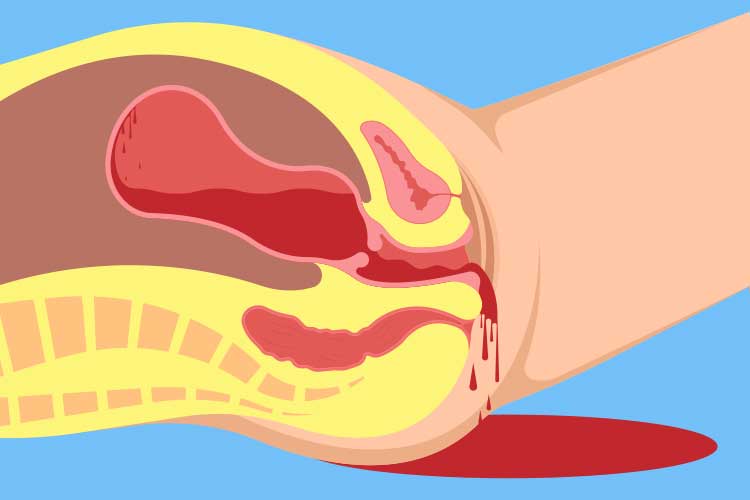
Postpartum Hemorrhage Causes Diagnosis and Management
## **Postpartum Hemorrhage Clinical Guide Causes Diagnosis and Management** ### **Definition** **Postpartum hemorrhage (PPH)** is excessive bleeding after childbirth. * **Primary (early) PPH:** ≥500 mL after vaginal delivery or ≥1000 mL after cesarean section within **24 hours** * **Secondary (late) PPH:** Excessive bleeding from **24 hours to 6 weeks** postpartum --- ## **Epidemiology and Importance** * Leading cause of **maternal mortality worldwide** * Rapid onset and progression require **early recognition and protocol-based management** --- ## **Pathophysiology** Normal hemostasis after delivery depends on **uterine contraction** compressing spiral arteries. Failure of contraction or disruption of clotting leads to uncontrolled bleeding. --- ## **Causes – “4 Ts” Framework** 1. **Tone (most common – uterine atony)** * Overdistended uterus (multiple pregnancy, polyhydramnios, macrosomia) * Prolonged or precipitous labor * Chorioamnionitis 2. **Trauma** * Cervical, vaginal, perineal tears * Uterine rupture * Hematomas 3. **Tissue** * Retained placental tissue * Placenta accreta spectrum 4. **Thrombin** * Coagulopathies (DIC, severe preeclampsia, HELLP, anticoagulant use) --- ## **Risk Factors** * Previous PPH * Operative delivery * Induction or augmentation of labor * Anemia * Placenta previa or accreta --- ## **Clinical Features** * Excessive vaginal bleeding * Boggy or enlarged uterus * Signs of hypovolemia: tachycardia, hypotension, pallor, altered sensorium * Reduced urine output --- ## **Initial Assessment and Diagnosis** **Diagnosis is clinical and urgent** * Quantify blood loss (visual + weighing) * Assess uterine tone * Inspect birth canal * Evaluate placenta completeness ### **Investigations (do not delay treatment)** * CBC (Hb, platelets) * Blood group and cross-match * Coagulation profile (PT, aPTT, fibrinogen) * ABG if severe shock --- ## **Management – Stepwise Approach** ### **Immediate Resuscitation** * Call for help * Airway and oxygen * Two wide-bore IV lines * Crystalloids followed by blood products (1:1:1 PRBC:plasma:platelets if massive) --- ## **Uterotonic Drugs (Cornerstone of Treatment)** ### **1. Oxytocin** * **Indication:** First-line for uterine atony * **Mechanism:** Stimulates uterine smooth muscle contraction * **Dose:** * IV infusion: 10–40 IU in 1 L NS/RL * IM: 10 IU * **Adverse effects:** Hypotension (rapid IV), water intoxication * **Contraindications:** None significant in PPH * **Monitoring:** Uterine tone, vitals * **Counselling:** First-line and safe --- ### **2. Methylergometrine** * **Mechanism:** Sustained uterine contraction via alpha-adrenergic stimulation * **Dose:** 0.2 mg IM (may repeat) * **Adverse effects:** Hypertension, nausea * **Contraindications:** Hypertension, preeclampsia, cardiac disease * **Monitoring:** Blood pressure --- ### **3. Carboprost (15-methyl PGF2α)** * **Mechanism:** Prostaglandin-induced myometrial contraction * **Dose:** 250 µg IM every 15–90 min (max 8 doses) * **Adverse effects:** Bronchospasm, diarrhea, fever * **Contraindications:** Asthma * **Monitoring:** Respiratory status --- ### **4. Misoprostol** * **Mechanism:** Prostaglandin E1 analog * **Dose:** 800–1000 µg rectal or sublingual * **Adverse effects:** Fever, shivering * **Use:** Low-resource settings --- ### **5. Tranexamic Acid** * **Indication:** All PPH within 3 hours of onset * **Mechanism:** Inhibits fibrinolysis * **Dose:** 1 g IV over 10 min (repeat once if bleeding continues) * **Adverse effects:** Rare thrombosis * **Contraindications:** Active thromboembolic disease * **Monitoring:** Renal function if repeated * **Counselling:** Reduces mortality when given early --- ## **Mechanical and Surgical Measures** ### **Mechanical** * Bimanual uterine massage * Uterine balloon tamponade (Bakri balloon) * Uterine packing ### **Surgical** * Uterine compression sutures (B-Lynch) * Uterine artery ligation * Internal iliac artery ligation * **Hysterectomy** (life-saving last resort) --- ## **Management by Cause** * **Atony:** Uterotonics → balloon → surgery * **Trauma:** Immediate repair of tears * **Tissue:** Manual removal, curettage * **Thrombin:** Correct coagulopathy with blood products --- ## **Secondary Postpartum Hemorrhage** **Causes** * Retained products * Subinvolution of uterus * Endometritis **Management** * Antibiotics * Uterotonics * Ultrasound-guided evacuation if indicated --- ## **Complications** * Hypovolemic shock * Acute kidney injury * DIC * Sheehan syndrome * Maternal death --- ## **Prevention** * Active management of third stage of labor * Antenatal anemia correction * Risk stratification and preparedness --- ## **Prognosis** Excellent with early recognition and protocol-driven care; delays increase morbidity and mortality. --- ## **SEO Meta Data** **SEO Title:** Postpartum Hemorrhage Causes Diagnosis and Management **Meta Description:** Comprehensive clinical guide on postpartum hemorrhage covering causes, diagnosis, stepwise management, uterotonic drugs, surgical options, prevention, and complications. **SEO Keywords (comma separated):** postpartum hemorrhage, PPH management, uterine atony, causes of PPH, tranexamic acid PPH, obstetric emergency, maternal hemorrhage, third stage labor complications --- ## **Frequently Asked Questions** **What is the most common cause of postpartum hemorrhage?** Uterine atony. **When should tranexamic acid be given in PPH?** Within 3 hours of onset of bleeding. **What is the first-line drug for PPH?** Oxytocin. **When is hysterectomy indicated in PPH?** When bleeding is uncontrollable and life-threatening despite conservative measures. **Can PPH occur after 24 hours?** Yes, it is termed secondary postpartum hemorrhage. --- If you want, I can **convert this into a CMS-ready HTML or PHP page**, **add FAQ schema and Article schema**, or **create MCQs and case-based questions** for your medical knowledge platform.
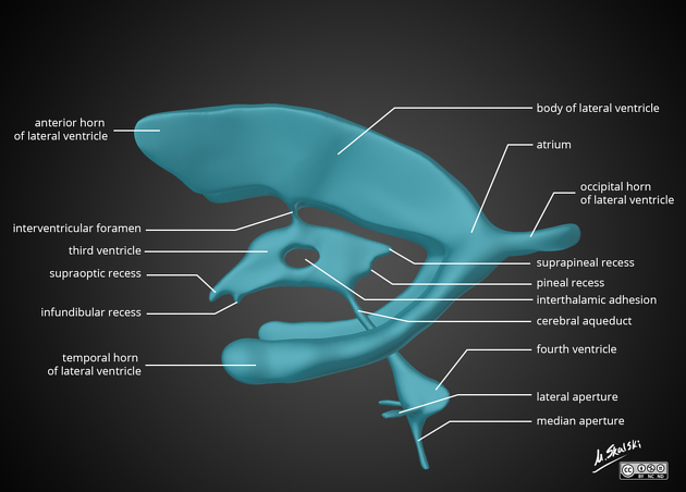
Cerebrospinal Fluid and Ventricular System Anatomy
## Cerebrospinal Fluid and Ventricular System Anatomy – Complete SEO-Friendly Guide ### SEO Title **Cerebrospinal Fluid and Ventricular System Anatomy** ### Meta Description Detailed anatomy of cerebrospinal fluid and the ventricular system covering formation, circulation, absorption, functions, ventricular components, and important clinical correlations. ### Keywords cerebrospinal fluid anatomy, ventricular system brain, lateral ventricles anatomy, third ventricle anatomy, fourth ventricle anatomy, CSF circulation, choroid plexus, arachnoid villi, hydrocephalus anatomy --- ## 1. Cerebrospinal Fluid (CSF) ### Definition Cerebrospinal fluid is a **clear, colorless fluid** that circulates within the **ventricular system of the brain and subarachnoid space** surrounding the brain and spinal cord, providing protection, nutrition, and waste removal. ### Normal Volume and Pressure * Total volume (adult): **≈150 mL** * Daily production: **≈500 mL** * Normal opening pressure (lumbar puncture): **70–180 mm H₂O** --- ## 2. Formation of CSF ### Choroid Plexus CSF is primarily produced by the **choroid plexus**, a vascular structure lined by **ependymal cells**. **Locations of choroid plexus** * Lateral ventricles (body and temporal horn) * Third ventricle * Fourth ventricle **Mechanism** * Active secretion via **Na⁺/K⁺ ATPase** * Water follows osmotically * Independent of intracranial pressure --- ## 3. Ventricular System of the Brain The ventricular system consists of **four interconnected cavities** lined by ependyma and filled with CSF. --- ### 3.1 Lateral Ventricles (First and Second Ventricles) **Location** * One in each cerebral hemisphere **Parts** 1. **Anterior (frontal) horn** * In frontal lobe * Roof: Corpus callosum * Floor: Head of caudate nucleus 2. **Body** * Extends through parietal lobe 3. **Posterior (occipital) horn** * In occipital lobe 4. **Inferior (temporal) horn** * In temporal lobe * Floor: Hippocampus * Roof: Tail of caudate nucleus **Communication** * Each lateral ventricle communicates with the third ventricle via the **interventricular foramen (foramen of Monro)** --- ### 3.2 Third Ventricle **Location** * Midline cavity between the two thalami **Boundaries** * Lateral walls: Thalamus and hypothalamus * Floor: Hypothalamus * Roof: Tela choroidea * Anterior wall: Lamina terminalis * Posterior wall: Pineal region **Connections** * Receives CSF from lateral ventricles * Drains into the fourth ventricle via the **cerebral aqueduct (aqueduct of Sylvius)** --- ### 3.3 Fourth Ventricle **Location** * Between pons and medulla anteriorly * Cerebellum posteriorly **Boundaries** * Floor: Rhomboid fossa * Roof: Superior and inferior medullary vela **Openings** * **One median aperture (foramen of Magendie)** * **Two lateral apertures (foramina of Luschka)** These openings allow CSF to enter the **subarachnoid space**. --- ## 4. Circulation of CSF **Flow pathway** 1. Lateral ventricles 2. Foramen of Monro 3. Third ventricle 4. Cerebral aqueduct 5. Fourth ventricle 6. Foramen of Magendie and Luschka 7. Subarachnoid space 8. Arachnoid villi and granulations 9. Superior sagittal sinus --- ## 5. Absorption of CSF ### Arachnoid Villi and Granulations * Protrusions of arachnoid mater into venous sinuses * Act as **one-way valves** * Absorption occurs when CSF pressure exceeds venous pressure Minor absorption also occurs via: * Spinal nerve sheaths * Choroid plexus --- ## 6. Composition of CSF * Clear and acellular * Low protein * Low potassium and calcium * Higher chloride compared to plasma * Glucose ≈ 60% of plasma glucose --- ## 7. Functions of CSF * **Mechanical protection** (shock absorber) * **Buoyancy** (reduces effective brain weight) * **Nutrient delivery** * **Removal of metabolic waste** * **Maintenance of intracranial pressure** --- ## 8. Blood–CSF Barrier Formed by: * Tight junctions between **choroid plexus epithelial cells** Functions: * Regulates composition of CSF * Protects CNS from toxins --- ## 9. Clinical Correlations ### Hydrocephalus * Abnormal accumulation of CSF **Types** * **Non-communicating (obstructive):** Block within ventricular system (e.g., aqueductal stenosis) * **Communicating:** Impaired absorption at arachnoid villi * **Normal pressure hydrocephalus:** Triad of gait disturbance, dementia, urinary incontinence --- ### Raised Intracranial Pressure * Headache * Vomiting * Papilledema * Altered consciousness --- ### Lumbar Puncture * Performed at **L3–L4 or L4–L5** * Measures CSF pressure and composition --- ## 10. High-Yield Exam Points * CSF production: **Choroid plexus** * Narrowest part of ventricular system: **Cerebral aqueduct** * Largest ventricles: **Lateral ventricles** * Main absorption site: **Arachnoid granulations** * CSF volume remains constant despite high daily production --- If you want, I can also provide **MCQs (exam-oriented)**, **clinical case-based questions**, or **schema-style revision tables** for CSF and ventricles.
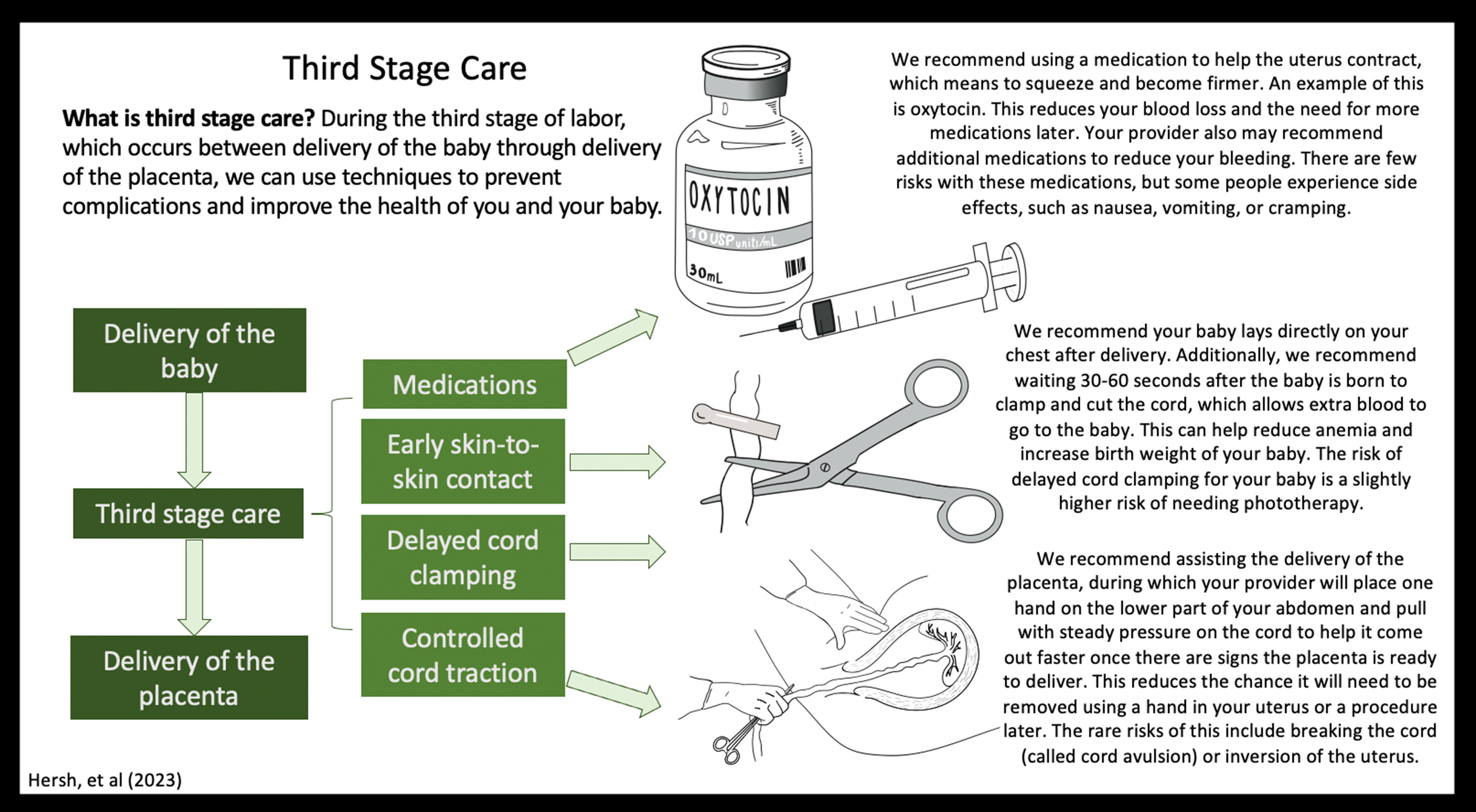
Active Management of Third Stage of Labor Complete Guide for PPH Prevention
Below is a **concise yet complete, exam-oriented, SEO-friendly reference** on **Active Management of Third Stage of Labor (AMTSL)**, structured for **medical students, clinicians, and obstetric exams**. --- # **Active Management of Third Stage of Labor (AMTSL)** ## **Definition** **Active Management of the Third Stage of Labor (AMTSL)** is a **planned set of interventions** performed **immediately after the birth of the baby** to **facilitate placental delivery and prevent postpartum hemorrhage (PPH)**, the leading cause of maternal mortality worldwide. --- ## **Importance and Rationale** * **Postpartum hemorrhage** accounts for a significant proportion of **maternal deaths** * AMTSL **reduces blood loss**, **shortens third stage**, and **lowers risk of uterine atony** * Recommended by **WHO, FIGO, ICM, and ACOG** --- ## **Components of AMTSL (Core Steps)** ### **1. Administration of a Uterotonic Drug (Most Important Step)** * Given **within 1 minute after delivery of the baby** * **After delivery of anterior shoulder or complete birth** * **Before or after placental delivery** (as per guideline) ### **2. Controlled Cord Traction (CCT)** * Gentle traction on umbilical cord * Combined with **counter-traction on uterus** * Performed **only after signs of placental separation** ### **3. Uterine Massage After Placental Delivery** * Ensures **uterine contraction** * Reduces risk of uterine atony * Routine sustained massage **not recommended**, but **assessment of tone is essential** --- ## **Uterotonic Drugs Used in AMTSL** ### **Oxytocin (Drug of Choice)** * **Dose:** 10 IU IM or slow IV * **Onset:** 2–3 minutes * **Advantages:** Highly effective, minimal side effects * **Preferred by WHO** ### **Ergometrine / Methylergometrine** * **Dose:** 0.2 mg IM/IV * **Contraindications:** Hypertension, pre-eclampsia, heart disease * Causes sustained uterine contraction ### **Oxytocin + Ergometrine (Syntometrine)** * More effective but **higher side effects** * Nausea, vomiting, hypertension ### **Misoprostol** * **Dose:** 600 μg orally * Used where injectables unavailable * Side effects: Fever, shivering --- ## **Controlled Cord Traction (CCT): Key Points** * Perform only when uterus is **well contracted** * Look for **signs of placental separation**: * Lengthening of cord * Gush of blood * Uterus becomes globular and rises * Prevents **retained placenta** * Reduces duration of third stage --- ## **Uterine Massage** * After placenta delivery * Assess uterine tone **every 15 minutes for first 2 hours** * Continuous massage is **not routinely advised** --- ## **Timing of Cord Clamping** * **Delayed cord clamping (1–3 minutes)** recommended * Does **not interfere with AMTSL** * Improves neonatal iron stores --- ## **Benefits of AMTSL** * ↓ Postpartum hemorrhage by **50–70%** * ↓ Severe blood loss (>1000 mL) * ↓ Need for blood transfusion * ↓ Duration of third stage * ↓ Maternal morbidity and mortality --- ## **AMTSL vs Expectant Management** | Feature | AMTSL | Expectant Management | | ------------- | ------------------ | -------------------- | | Uterotonic | Given routinely | Not given | | Cord traction | Yes | No | | Blood loss | Significantly less | More | | Duration | Shorter | Longer | | PPH risk | Low | High | --- ## **Indications** * **All vaginal deliveries** * Especially important in: * Anemia * Multiple pregnancy * Prolonged labor * Polyhydramnios * Grand multiparity --- ## **Contraindications** * No absolute contraindication * Avoid **ergometrine** in: * Hypertension * Preeclampsia * Cardiac disease --- ## **Complications (Rare)** * Retained placenta (if CCT done improperly) * Uterine inversion (improper traction) * Drug side effects (ergometrine, misoprostol) --- ## **WHO Recommended AMTSL Protocol (Summary)** 1. **Oxytocin 10 IU IM/IV** 2. **Delayed cord clamping** 3. **Controlled cord traction** 4. **Assess uterine tone** --- ## **Key Exam Pearls** * **Most important component:** Uterotonic administration * **Drug of choice:** Oxytocin * **Primary aim:** Prevention of postpartum hemorrhage * **Third stage normal duration:** ≤ 30 minutes --- ## **Conclusion** **Active Management of Third Stage of Labor** is a **simple, cost-effective, life-saving obstetric intervention**. Universal implementation of AMTSL significantly reduces **postpartum hemorrhage and maternal mortality**, making it a **standard of care in modern obstetrics**. --- ### If you want next: * **SEO title and meta keywords** * **High-yield MCQs (exam mode) in single HTML-CSS-JS file** * **Clinical flowchart image** * **Comparison table for exams** Just tell me 👍
Induction of Labor Indications Methods Contraindications Complications
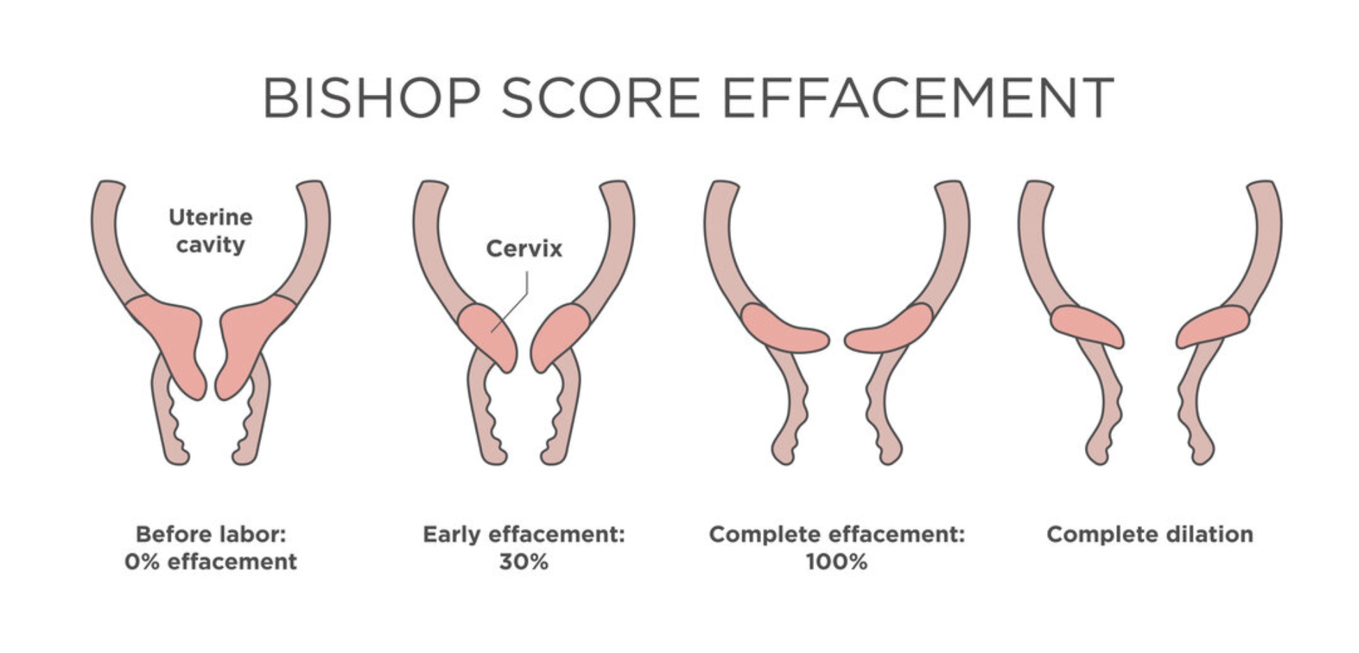
Below is a **concise yet complete, SEO-friendly, exam-ready reference** on **Induction of Labor**, structured with clear headings (H1–H3 style) and high-yield clinical content. --- # **Induction of Labor** ## **Definition** **Induction of labor (IOL)** is the **artificial initiation of uterine contractions** before the spontaneous onset of labor, with the aim of achieving **vaginal delivery**, when continuation of pregnancy poses **greater maternal or fetal risk** than delivery. --- ## **Indications for Induction of Labor** ### **Maternal Indications** * Post-term pregnancy (≥41 weeks) * Pre-eclampsia and eclampsia * Gestational hypertension * Diabetes mellitus (gestational or pre-existing) * Premature rupture of membranes (PROM) at term * Chorioamnionitis * Rh isoimmunization * Maternal medical disorders (renal, cardiac disease) * Intrauterine fetal demise (IUFD) ### **Fetal Indications** * Intrauterine growth restriction (IUGR) * Oligohydramnios * Non-reassuring fetal status (controlled setting) * Fetal anomalies requiring early delivery ### **Elective Induction** * At ≥39 weeks with confirmed gestational age and favorable cervix --- ## **Contraindications to Induction of Labor** ### **Absolute Contraindications** * Placenta previa * Vasa previa * Transverse lie * Cord prolapse * Previous classical cesarean section * Previous uterine rupture * Invasive cervical cancer * Cephalopelvic disproportion (CPD) ### **Relative Contraindications** * Multiple previous cesarean sections * Grand multiparity * Unstable lie * Active genital herpes --- ## **Pre-Induction Assessment** Show readiness of cervix and maternal-fetal safety. ### **Bishop Score** Assesses **cervical favorability** using: * Cervical dilatation * Effacement * Consistency * Position * Fetal station **Score interpretation:** * **≥6–8** → Favorable cervix (high success) * **<6** → Unfavorable cervix (requires cervical ripening) --- ## **Methods of Induction of Labor** ### **1. Mechanical Methods** * **Membrane sweeping** * **Foley catheter (balloon catheter)** * Double balloon catheter **Advantages:** Low cost, minimal uterine hyperstimulation **Disadvantages:** Discomfort, infection risk --- ### **2. Pharmacological Methods** #### **Prostaglandins** * **Dinoprostone (PGE₂)** – Gel, tablet, vaginal insert * **Misoprostol (PGE₁)** – Oral or vaginal **Actions:** * Cervical ripening * Uterine contractions **Contraindications:** * Previous uterine scar (relative for misoprostol) * Fetal distress * Hypersensitivity --- #### **Oxytocin** * Used after cervical ripening * IV infusion with titration **Mechanism:** Stimulates uterine smooth muscle contraction **Monitoring:** Continuous CTG required --- ### **3. Surgical Methods** * **Amniotomy (Artificial rupture of membranes)** **Prerequisites:** * Engaged head * Favorable cervix * No placenta previa --- ## **Monitoring During Induction** * Continuous fetal heart rate monitoring * Maternal vitals * Uterine contraction pattern * Progress of labor (partograph) --- ## **Complications of Induction of Labor** ### **Maternal Complications** * Uterine hyperstimulation * Uterine rupture * Postpartum hemorrhage * Infection * Failed induction leading to cesarean section ### **Fetal Complications** * Fetal distress * Meconium aspiration * Birth asphyxia * Cord prolapse (after amniotomy) --- ## **Failed Induction** **Definition:** Failure to establish active labor after adequate attempts with appropriate methods. **Management:** * Re-assess indication * Repeat cervical ripening * Cesarean delivery if indicated --- ## **Key Clinical Pearls** * Always confirm **gestational age** before induction * Bishop score guides method selection * Avoid prostaglandins in scarred uterus (especially misoprostol) * Continuous monitoring is mandatory * Individualize induction based on maternal and fetal condition --- ## **Conclusion** Induction of labor is a **critical obstetric intervention** that improves maternal and fetal outcomes when appropriately indicated. Proper **case selection, cervical assessment, method choice, and vigilant monitoring** are essential to maximize success and minimize complications. --- If you want, I can next provide: * **SEO title, meta description, and keywords** * **Exam-oriented tables** * **Case-based MCQs (HTML + CSS + JS, single file, modern UI)** * **Flowcharts for induction decision-making**
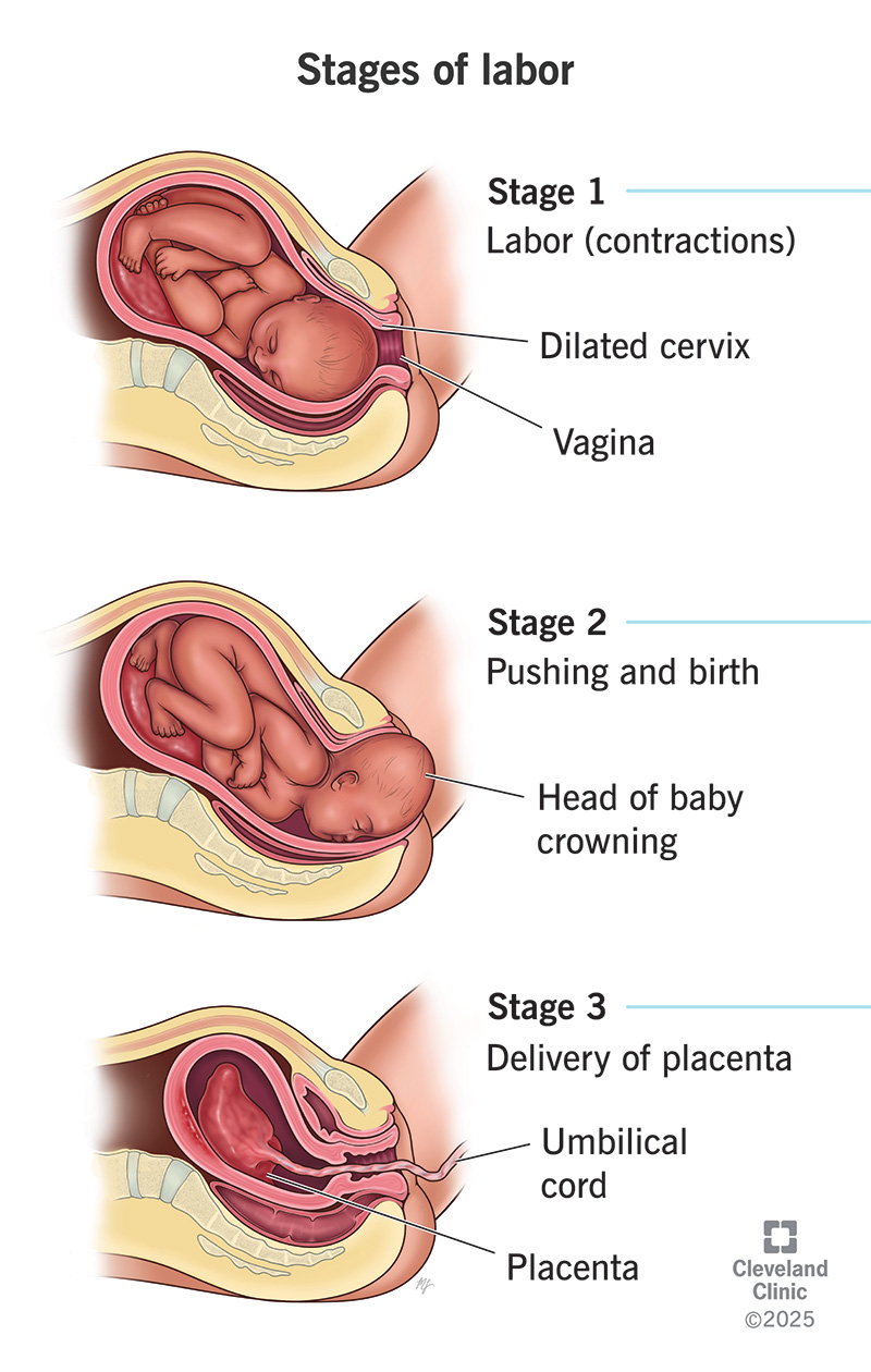
Normal Labour Definition Stages Physiology Management and Clinical Features
Below is a **concise yet complete, SEO-friendly medical reference** on **Normal Labour**, structured with clear **H1–H3 headings** and exam-oriented clarity. --- # **Normal Labour** ## **Definition of Normal Labour** Normal labour is a **physiological process** in which a **term pregnancy (37–42 weeks)** culminates in the **spontaneous onset of labour**, resulting in **vaginal delivery of a single, live fetus in vertex presentation**, followed by expulsion of the placenta, **without maternal or fetal complications**. --- ## **Criteria of Normal Labour** Normal labour fulfills all of the following: * **Gestational age:** 37–42 weeks * **Onset:** Spontaneous * **Presentation:** Vertex (cephalic) * **Number of fetus:** Singleton * **Progress:** Regular uterine contractions with progressive cervical dilatation * **Mode of delivery:** Vaginal, without operative intervention * **Outcome:** Healthy mother and baby --- ## **Physiology of Normal Labour** Labour is initiated by a complex interaction of: * **Hormonal factors:** ↑ Estrogen, ↓ Progesterone dominance, ↑ Prostaglandins, ↑ Oxytocin receptors * **Uterine activity:** Coordinated, rhythmic contractions starting from fundus * **Cervical changes:** Effacement and dilatation due to collagen remodeling * **Fetal contribution:** Fetal HPA axis activation and cortisol release --- ## **Stages of Normal Labour** ### **First Stage of Labour** **From onset of true labour pains to full cervical dilatation (10 cm)** #### Latent Phase * Cervical dilatation: 0–3/4 cm * Contractions: Mild, irregular * Duration: * Primigravida: up to 8–12 hours * Multigravida: shorter #### Active Phase * Cervical dilatation: 4–10 cm * Contractions: Regular, strong, 3–5 per 10 minutes * Rate of dilatation: * Primigravida: ~1 cm/hour * Multigravida: ~1.5 cm/hour --- ### **Second Stage of Labour** **From full dilatation to delivery of the fetus** * Duration: * Primigravida: ≤2 hours (≤3 hours with epidural) * Multigravida: ≤1 hour * Mechanism of labour occurs: * Engagement * Descent * Flexion * Internal rotation * Extension * Restitution and external rotation * Expulsion --- ### **Third Stage of Labour** **From delivery of fetus to expulsion of placenta** * Duration: ≤30 minutes * Placental separation mechanisms: * Schultze (central separation) * Duncan (marginal separation) * Managed actively to prevent postpartum hemorrhage --- ## **Mechanism of Normal Labour (Vertex Presentation)** 1. Engagement 2. Descent 3. Flexion 4. Internal rotation 5. Extension 6. Restitution 7. External rotation 8. Expulsion --- ## **Signs of True Labour** * Regular, painful uterine contractions * Progressive cervical effacement and dilatation * Show (blood-stained mucus) * Descent of presenting part --- ## **Monitoring in Normal Labour** * **Maternal:** Pulse, BP, temperature, uterine contractions * **Fetal:** Fetal heart rate monitoring * **Progress:** Partograph assessment of cervical dilatation, descent, and contractions --- ## **Management Principles of Normal Labour** * Supportive care and reassurance * Adequate hydration and nutrition * Pain relief (non-pharmacologic or pharmacologic) * Asepsis and bladder care * Continuous monitoring for deviations from normal --- ## **Outcome of Normal Labour** * Vaginal birth without complications * Minimal maternal morbidity * Healthy neonate with good Apgar score --- ## **Clinical Importance** * Benchmark for identifying **abnormal labour patterns** * Essential for **obstetric exams**, labour ward practice, and safe maternal care * Basis for using **partograph** and deciding interventions --- If you want, I can next provide: * **SEO title, meta description, and keywords** * **Abnormal labour comparison** * **Partograph-based exam notes** * **MCQs or case-based questions in HTML/CSS/JS (single file, modern UI)**
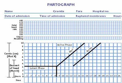
Partogram in Obstetrics Complete Guide for Monitoring Labour Progress
Below is a **single-place, exam-ready, clinically complete, SEO-friendly reference** on **Partogram**, written in a **clear H1–H4 structure** suitable for medical students, nurses, midwives, and clinicians. --- # **Partogram in Obstetrics: Complete Guide for Monitoring Labour Progress** ## **Introduction to Partogram** A **partogram (partograph)** is a **graphical tool** used to **monitor the progress of labour**, assess **maternal and fetal well-being**, and guide **timely clinical interventions**. It is a **WHO-recommended, low-cost, high-impact tool** for preventing **prolonged and obstructed labour**, thereby reducing **maternal and perinatal morbidity and mortality**. --- ## **Definition of Partogram** A **partogram** is a **chart that plots cervical dilatation against time**, along with **uterine contractions, fetal condition, and maternal parameters**, to provide a **continuous visual record of labour**. --- ## **Objectives of Using a Partogram** * Detect **abnormal labour progress early** * Identify **prolonged labour** * Prevent **obstructed labour** * Reduce **maternal complications** (PPH, sepsis, uterine rupture) * Reduce **fetal complications** (birth asphyxia, stillbirth) * Assist in **decision-making and referral** --- ## **Types of Partogram** ### **1. WHO Modified Partogram** * **Active phase starts at 4 cm cervical dilatation** * **Latent phase excluded** * Most commonly used in clinical practice ### **2. WHO Composite Partogram (Older)** * Includes **latent and active phase** * Latent phase up to **8 hours** * Less commonly used now ### **3. Simplified Partogram** * Focuses on **key parameters only** * Used in **low-resource settings** --- ## **Components of a Partogram** ### **A. Fetal Condition** Monitored to assess fetal well-being. #### **1. Fetal Heart Rate (FHR)** * Recorded **every 30 minutes** * Normal: **110–160 beats/min** * Abnormal: * <110 → Bradycardia * > 160 → Tachycardia #### **2. Amniotic Fluid** * **I** – Intact membranes * **C** – Clear liquor * **M** – Meconium-stained * **B** – Blood-stained #### **3. Moulding of Fetal Skull** * 0 → Sutures separated * * → Sutures touching * ++ → Sutures overlapping (reducible) * +++ → Sutures overlapping (irreducible) → **danger sign** --- ### **B. Progress of Labour** #### **1. Cervical Dilatation** * Plotted with **X** * Expected rate in active phase: **≥1 cm/hour** * Starts at **4 cm** #### **2. Descent of Head** * Plotted with **O** * Measured in **fifths palpable abdominally** * Or by **station (–5 to +5)** --- ### **C. Uterine Contractions** Recorded **every 30 minutes**: | Number / 10 min | Interpretation | | --------------- | ---------------- | | <2 | Inadequate | | 3–4 | Adequate | | ≥5 | Hyperstimulation | **Duration**: * <20 sec – Mild * 20–40 sec – Moderate * > 40 sec – Strong --- ### **D. Alert Line and Action Line** #### **Alert Line** * Drawn from **4 cm to full dilatation at 1 cm/hour** * Labour to the **left** → Normal * Crossing → **Slow progress** #### **Action Line** * Drawn **4 hours to the right of alert line** * Crossing → **Immediate intervention required** --- ### **E. Maternal Condition** #### **1. Pulse** * Every **30 minutes** #### **2. Blood Pressure** * Every **4 hours** * More frequent if abnormal #### **3. Temperature** * Every **2 hours** #### **4. Urine Examination** * Volume * Protein * Ketones --- ## **Interpretation of Partogram** ### **Normal Labour** * Cervical dilatation remains **left of alert line** * Adequate contractions * Normal fetal heart rate ### **Prolonged Labour** * Cervical dilatation crosses **alert line** * Requires: * Re-assessment * Amniotomy * Oxytocin augmentation * Referral if needed ### **Obstructed Labour** * Cervical dilatation reaches **action line** * Features: * No descent of head * Severe moulding (+++) * Maternal exhaustion * Management: * Operative delivery (CS / instrumental) --- ## **Indications for Using a Partogram** * All women in **active labour** * Especially useful in: * Primigravida * Induced labour * Previous prolonged labour * Referral cases --- ## **Contraindications / Limitations** * Not used in **latent phase (<4 cm)** * Less effective without **trained staff** * Requires **regular monitoring** --- ## **Advantages of Partogram** * Simple and cost-effective * Visual and easy to interpret * Reduces: * Prolonged labour * Obstructed labour * Unnecessary interventions * Improves maternal and neonatal outcomes --- ## **Disadvantages** * Incorrect plotting may mislead decisions * Requires training and adherence * Limited use without timely action --- ## **Role of Partogram in Modern Obstetrics** * Key tool in **Safe Motherhood Initiative** * Recommended by **WHO, FIGO** * Essential in **institutional deliveries** * Supports **evidence-based labour management** --- ## **Exam-Oriented Clinical Pearls** * **Alert line crossing** → Observe and evaluate * **Action line crossing** → Act immediately * **Moulding +++** → Suggests CPD * **Meconium liquor** → Fetal distress * **Rate <1 cm/hr** → Prolonged labour --- ## **Conclusion** The **partogram is a cornerstone of intrapartum care**, enabling **early detection of abnormal labour**, guiding **timely interventions**, and significantly improving **maternal and neonatal outcomes**. Proper understanding and correct use of the partogram are essential skills for all healthcare providers involved in childbirth. --- If you want next: * **SEO title, meta description, and keywords** * **Image-based explanation** * **25 hard case-based MCQs** * **HTML CSS JS interactive partogram simulator** Just tell me 👍
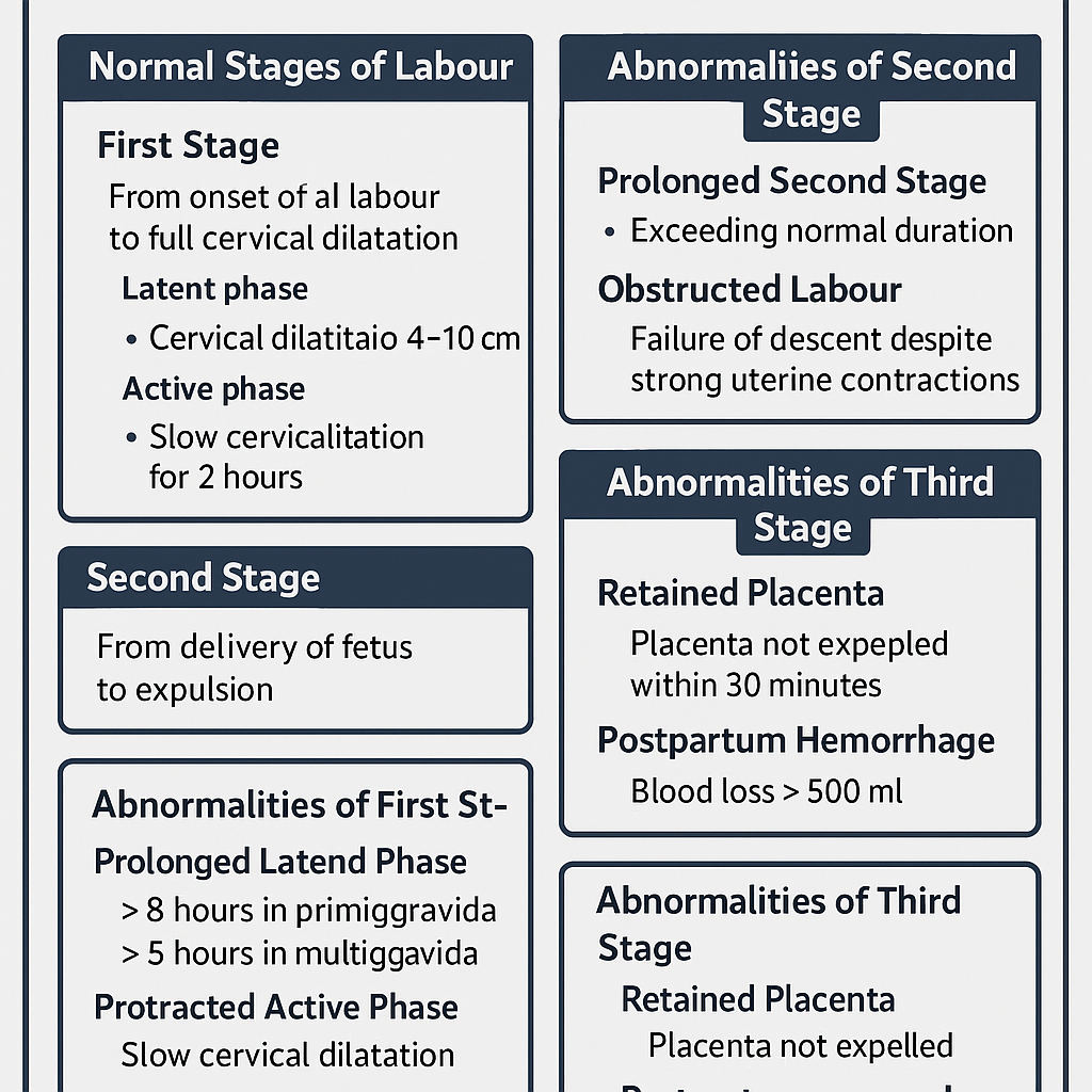
Stages of Labour Normal and Abnormal Progress Complete Clinical Guide
## **Stages of Labour Normal and Abnormal Progress Complete Clinical Guide** ### **SEO Description** Comprehensive obstetrics guide explaining normal and abnormal stages of labour with mechanisms, duration, clinical features, diagnosis, complications, and management for exams and clinical practice. ### **SEO Keywords** stages of labour, normal labour stages, abnormal labour, prolonged labour, obstructed labour, dysfunctional labour, first stage labour, second stage labour, third stage labour, active labour management, obstetrics labour guide --- # **Stages of Labour Normal and Abnormal** Labour is the **physiological process by which the fetus, placenta, and membranes are expelled from the uterus after fetal viability** through coordinated uterine contractions resulting in cervical effacement and dilatation. --- ## **Classification of Labour** * **Normal Labour**: Spontaneous onset, singleton, cephalic presentation, term pregnancy, no complications, vaginal delivery. * **Abnormal Labour**: Any deviation in onset, duration, progress, or outcome of labour. --- # **Normal Stages of Labour** ## **First Stage of Labour** **Definition:** Period from onset of true labour pains to full cervical dilatation (10 cm). ### **Phases** #### **Latent Phase** * Cervical dilatation: 0–3 cm * Cervical effacement occurs * Contractions mild to moderate * Duration: * Primigravida: up to 8 hours * Multigravida: up to 5 hours #### **Active Phase** * Cervical dilatation: 4–10 cm * Rapid dilatation * Strong, regular contractions * Rate of dilatation: * Primigravida: ~1 cm/hour * Multigravida: ~1.5 cm/hour ### **Normal Mechanism** * Uterine contractions * Cervical effacement and dilatation * Formation of forewaters and bulging membranes --- ## **Second Stage of Labour** **Definition:** Period from full cervical dilatation to delivery of the fetus. ### **Characteristics** * Strong expulsive uterine contractions * Voluntary maternal bearing down * Fetal descent, flexion, rotation, extension, restitution, expulsion ### **Duration** * Primigravida: up to 2 hours (3 hours with epidural) * Multigravida: up to 1 hour (2 hours with epidural) --- ## **Third Stage of Labour** **Definition:** Period from delivery of fetus to expulsion of placenta and membranes. ### **Mechanism** * Placental separation * Placental descent * Placental expulsion ### **Duration** * Usually within 5–15 minutes * Maximum acceptable: 30 minutes ### **Signs of Placental Separation** * Uterus becomes globular and firm * Lengthening of umbilical cord * Sudden gush of blood * Uterine fundus rises --- # **Abnormal Labour** Abnormal labour occurs due to problems related to **powers, passenger, or passage**. --- ## **Abnormalities of First Stage** ### **Prolonged Latent Phase** * > 8 hours in primigravida * > 5 hours in multigravida > **Causes** * Ineffective uterine contractions * Anxiety, dehydration * False labour **Management** * Reassurance * Hydration and analgesia * Rule out cephalopelvic disproportion --- ### **Protracted Active Phase** * Slow cervical dilatation **Causes** * Uterine inertia * Malposition * Mild CPD **Management** * Partograph monitoring * Amniotomy * Oxytocin augmentation if indicated --- ### **Arrest of Dilatation** * No cervical dilatation for 2 hours **Causes** * Obstructed labour * Malpresentation **Management** * Evaluate cause * Cesarean delivery if obstruction present --- ## **Abnormalities of Second Stage** ### **Prolonged Second Stage** * Exceeds normal duration **Causes** * Ineffective pushing * Epidural analgesia * Fetal malposition * Pelvic inadequacy **Complications** * Maternal exhaustion * Fetal distress **Management** * Assisted vaginal delivery * Cesarean section if indicated --- ### **Obstructed Labour** **Definition:** Failure of descent despite strong uterine contractions. **Causes** * Cephalopelvic disproportion * Fetal anomalies * Pelvic tumors **Complications** * Uterine rupture * Fetal hypoxia **Management** * Emergency cesarean section --- ## **Abnormalities of Third Stage** ### **Retained Placenta** * Placenta not expelled within 30 minutes **Types** * Placenta adherens * Trapped placenta * Placenta accreta spectrum **Management** * Active management * Manual removal under anesthesia --- ### **Postpartum Hemorrhage** * Blood loss >500 ml vaginal delivery **Causes** * Uterine atony * Retained placental tissue **Management** * Uterotonics * Bimanual compression * Surgical intervention if required --- # **Clinical Monitoring of Labour** * Partograph use * Fetal heart rate monitoring * Maternal vitals * Progress of cervical dilatation and descent --- # **Summary Table** | Stage | Normal Duration | Common Abnormalities | | ------ | --------------- | ---------------------- | | First | Latent + Active | Prolonged, Arrest | | Second | ≤2 hours | Prolonged, Obstructed | | Third | ≤30 minutes | Retained placenta, PPH | --- ## **Key Exam Pearls** * Active phase starts at 4 cm dilatation * Arrest disorders require prompt intervention * Third stage complications cause most maternal morbidity * Partograph is essential for early detection of abnormal labour --- If you want, I can **add MCQs, case based questions, management algorithms, or convert this into an exam-ready PDF or website content**.
Fetal Skull Anatomy in Obstetrics: Sutures, Fontanelles, Diameters and Clinical Importance
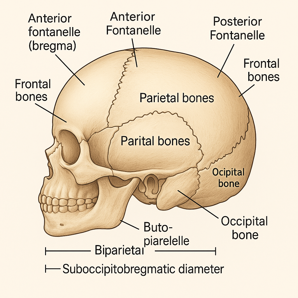
## **SEO Title** **Fetal Skull Anatomy in Obstetrics: Sutures, Fontanelles, Diameters and Clinical Importance** ## **Meta Description** Comprehensive obstetrics-focused guide on fetal skull anatomy covering bones, sutures, fontanelles, diameters, molding, and their clinical significance during labor and delivery. ## **SEO Keywords** fetal skull obstetrics, fetal skull anatomy, sutures of fetal skull, fontanelles fetal skull, fetal skull diameters, molding of fetal skull, obstetric diameters, fetal head engagement, labor mechanism fetal skull --- # **Fetal Skull – Complete Obstetric Guide** ## **Introduction** The **fetal skull** is the most important part of the fetus in obstetrics because its **size, shape, flexibility, and diameters** determine the **mechanism of labor**, engagement, descent, and mode of delivery. --- ## **1. Structure of the Fetal Skull** The fetal skull is divided into **three main parts**: ### **A. Vault (Cranium) – Obstetrically Most Important** * Formed by **membranous bones** * Flexible and compressible * Allows **molding during labor** **Bones of the vault:** * 2 Frontal bones * 2 Parietal bones * Upper part of occipital bone --- ### **B. Base of the Skull** * Formed by **cartilaginous bones** * Rigid and non-compressible * Not affected by molding **Bones include:** * Sphenoid * Temporal * Lower occipital bone --- ### **C. Face** * Small obstetric significance * Important in **face presentation** --- ## **2. Sutures of the Fetal Skull** **Sutures** are fibrous joints between skull bones that allow overlapping during labor. | Suture | Location | Obstetric Importance | | ---------------------------- | ---------------------------------- | ---------------------------- | | **Sagittal suture** | Between parietal bones | Determines head position | | **Coronal suture** | Between frontal & parietal bones | Identifies degree of flexion | | **Lambdoid suture** | Between parietal & occipital bones | Helps locate occiput | | **Frontal (metopic) suture** | Between frontal bones | Indicates head attitude | --- ## **3. Fontanelles** Fontanelles are **membranous gaps** at the junction of sutures. ### **A. Anterior Fontanelle (Bregma)** * Diamond-shaped * Junction of **sagittal, coronal, and frontal sutures** * Closes by **18 months** * Palpation indicates **deflexed head** ### **B. Posterior Fontanelle (Lambda)** * Triangular * Junction of **sagittal and lambdoid sutures** * Closes by **6–8 weeks** * Palpation indicates **well-flexed vertex** **👉 Posterior fontanelle is the key landmark in normal labor** --- ## **4. Diameters of the Fetal Skull** Diameters are classified into **transverse and anteroposterior**. ### **A. Transverse Diameters** | Diameter | Measurement | Importance | | ----------------------------- | ----------- | ----------------- | | **Biparietal diameter (BPD)** | **9.5 cm** | Engaging diameter | | Bitemporal | 8 cm | Internal fit | | Bimastoid | 7.5 cm | Base diameter | --- ### **B. Anteroposterior Diameters** | Diameter | Measurement | Presentation | | ------------------------ | ----------- | -------------------------- | | **Suboccipitobregmatic** | **9.5 cm** | Well-flexed vertex (ideal) | | Suboccipitofrontal | 10 cm | Incomplete flexion | | Occipitofrontal | 11.5 cm | Deflexed vertex | | Mentovertical | 13.5 cm | Brow presentation | | Submentobregmatic | 9.5 cm | Face presentation | --- ## **5. Circumferences of the Fetal Head** | Circumference | Measurement | | -------------------- | ----------- | | Suboccipitobregmatic | ~32 cm | | Occipitofrontal | ~34 cm | | Mentovertical | ~38 cm | --- ## **6. Molding of the Fetal Skull** **Molding** refers to **overlapping of skull bones during labor**. ### **Grades of Molding** * **+ (Mild):** Sutures touching * **++ (Moderate):** Sutures overlapping but reducible * **+++ (Severe):** Overlapping not reducible (suggests CPD) **Clinical Importance** * Facilitates vaginal delivery * Excessive molding → fetal distress or obstructed labor --- ## **7. Caput Succedaneum** * Edematous swelling of scalp * Crosses suture lines * Appears at birth * Resolves spontaneously --- ## **8. Cephalhematoma** * Subperiosteal hemorrhage * Does **not cross suture lines** * Appears hours after birth * Resolves slowly --- ## **9. Obstetric Importance of the Fetal Skull** * Determines **engagement** * Helps assess **presentation and position** * Influences **mechanism of labor** * Guides **decision for vaginal delivery vs cesarean** * Critical in diagnosing **CPD and malpresentations** --- ## **Key Clinical Points** * **Best diameter for vaginal delivery:** Suboccipitobregmatic (9.5 cm) * **Most dangerous presentation:** Brow (mentovertical 13.5 cm) * **Posterior fontanelle palpable → normal vertex** * **Excessive molding → obstructed labor** --- If you want, I can also provide: * **SEO FAQs** * **Exam-oriented tables** * **25 hard case-based MCQs in HTML/CSS/JS (single file, modern UI)** * **Illustration prompts for image generation** Just tell me 👍

Contracted Pelvis and Cephalopelvic Disproportion Types Diagnosis Management
Below is a **concise yet complete, exam-oriented and SEO-friendly medical reference** covering **Contracted Pelvis, Cephalopelvic Disproportion (CPD), and Types of Pelvis**. Structured with **H1–H4 headings**, keyword-rich language, and clear clinical flow. **No images included**, as requested. --- # Contracted Pelvis and Cephalopelvic Disproportion: Types, Diagnosis and Management ## Introduction **Contracted pelvis** and **cephalopelvic disproportion (CPD)** are major causes of **obstructed labor**, **prolonged labor**, and **operative delivery**. Understanding **pelvic anatomy**, **pelvic types**, and **fetal–pelvic relationships** is essential for safe obstetric practice. --- ## Contracted Pelvis ### Definition A **contracted pelvis** is a pelvis in which **one or more pelvic diameters are reduced** below normal, interfering with the **descent and delivery of the fetus**. --- ### Causes of Contracted Pelvis #### Developmental Causes * Congenital pelvic abnormalities * Malnutrition during childhood * Rickets and osteomalacia #### Acquired Causes * Pelvic fractures with malunion * Tuberculosis of bones * Spinal deformities (kyphosis, scoliosis) * Previous pelvic surgery --- ### Classification of Contracted Pelvis #### 1. Anatomical Contracted Pelvis * **True contraction** of one or more diameters * Diagnosed by **pelvimetry** #### 2. Functional Contracted Pelvis * Pelvic diameters normal * Obstruction due to: * Large fetus * Malposition or malpresentation * Deflexed fetal head --- ### Degrees of Contracted Pelvis (Based on Obstetric Conjugate) | Degree | Obstetric Conjugate | Clinical Significance | | -------- | ------------------- | ------------------------- | | Mild | 9–10 cm | Vaginal delivery possible | | Moderate | 7.5–9 cm | Trial of labor | | Severe | <7.5 cm | Cesarean section | --- ## Cephalopelvic Disproportion (CPD) ### Definition **Cephalopelvic disproportion** occurs when the **fetal head is too large** or the **maternal pelvis too small** to allow vaginal delivery. --- ### Types of CPD #### 1. Absolute CPD * Gross pelvic contraction * Vaginal delivery **impossible** #### 2. Relative CPD * Borderline pelvis * Delivery depends on: * Fetal head molding * Position and attitude * Strength of uterine contractions --- ### Causes of CPD #### Maternal Factors * Contracted pelvis * Pelvic tumors * Short stature #### Fetal Factors * Macrosomia * Hydrocephalus * Occipitoposterior position * Deflexed head --- ### Clinical Features of CPD * Failure of head engagement * Prolonged labor * Cervical dystocia * Increasing caput and molding * Maternal exhaustion * Fetal distress --- ### Diagnosis of CPD #### Antenatal Assessment * History of difficult labor * Clinical pelvimetry * Ultrasound for fetal weight #### Intrapartum Diagnosis * Lack of descent despite good contractions * Non-progress of labor * Rising Bandl’s ring --- ### Management of CPD #### Antenatal * Identify high-risk cases * Plan mode of delivery #### Intrapartum * **Trial of labor** in selected cases * Continuous maternal and fetal monitoring #### Definitive Management * **Cesarean section** for: * Absolute CPD * Failed trial of labor * Fetal distress --- ## Types of Pelvis (Caldwell–Moloy Classification) ### 1. Gynecoid Pelvis * Most favorable for vaginal delivery * Rounded inlet * Wide subpubic angle * Straight side walls --- ### 2. Android Pelvis * Male-type pelvis * Heart-shaped inlet * Narrow mid-pelvis * Common cause of **arrest of descent** --- ### 3. Anthropoid Pelvis * Oval inlet (anteroposterior diameter increased) * Occipitoposterior position common * Vaginal delivery usually possible --- ### 4. Platypelloid Pelvis * Flattened pelvis * Wide transverse diameter * Narrow anteroposterior diameter * Engagement delayed --- ## Comparison of Pelvic Types | Pelvic Type | Inlet Shape | Labor Outcome | | ------------ | ------------ | ------------------ | | Gynecoid | Round | Best | | Android | Heart-shaped | Difficult | | Anthropoid | Oval (AP) | OP common | | Platypelloid | Flat | Engagement delayed | --- ## Clinical Importance * Major determinant of **mode of delivery** * Prevention of **obstructed labor** * Reduces maternal and perinatal morbidity * Essential for **exam preparation** and **clinical decision-making** --- ## Conclusion **Contracted pelvis and CPD** remain critical challenges in obstetrics. Accurate **pelvic assessment**, understanding **pelvic types**, and timely intervention ensure safe outcomes for both mother and fetus. --- ### SEO Keywords (Comma-Separated) contracted pelvis, cephalopelvic disproportion, CPD in obstetrics, types of pelvis, gynecoid pelvis, android pelvis, anthropoid pelvis, platypelloid pelvis, obstructed labor causes, pelvic contraction degrees, CPD diagnosis management If you want, I can also provide **case-based MCQs**, **exam notes**, or a **single-file HTML CSS JS quiz** on this topic.
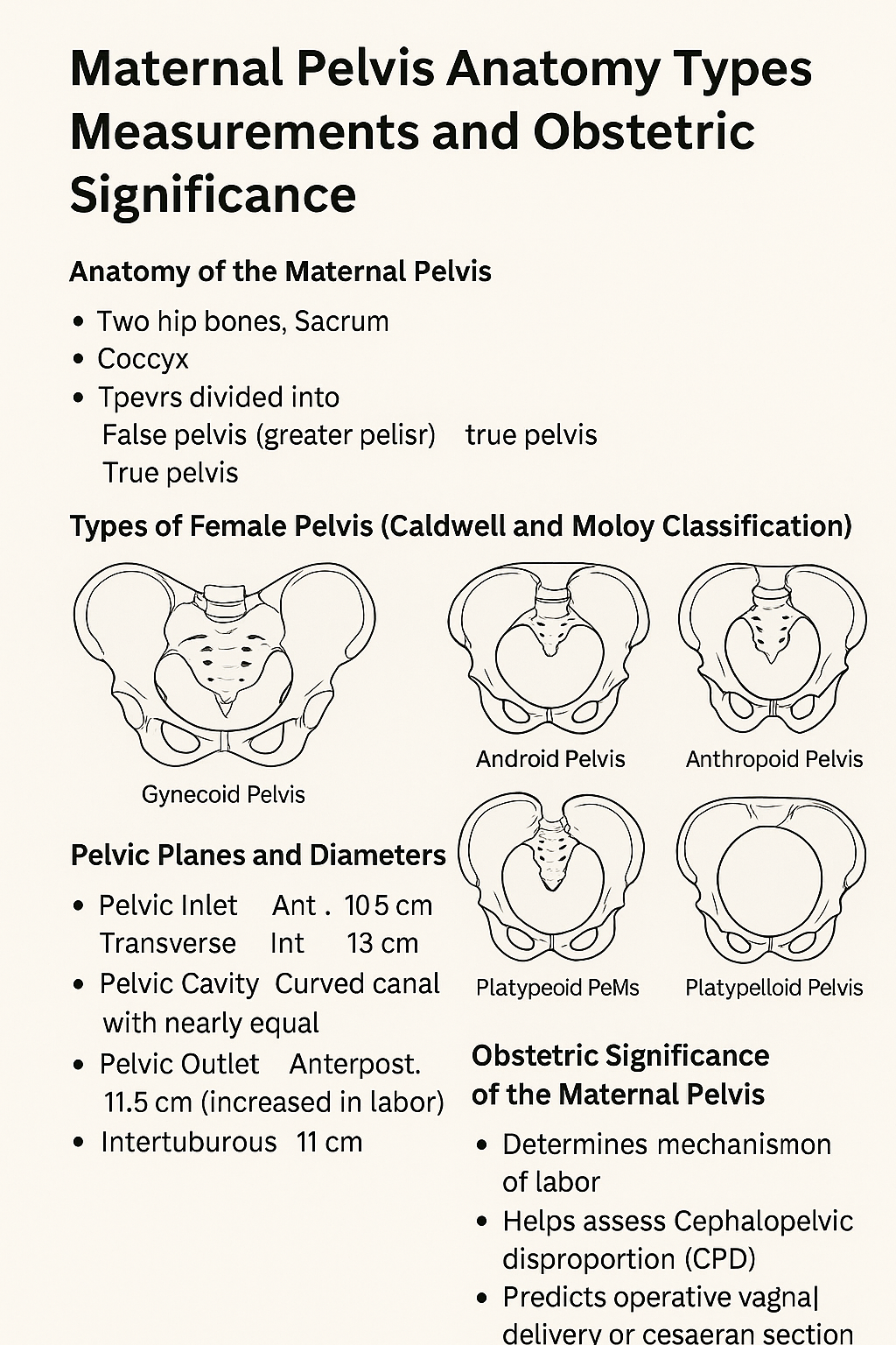
Maternal Pelvis Anatomy Types Measurements and Obstetric Significance
# **Maternal Pelvis Anatomy Types Measurements and Obstetric Significance** ## **Introduction** The **maternal pelvis** is a rigid bony structure that forms the birth canal. Its **shape, size, and dimensions** are critical determinants of **labor progression, fetal descent, and delivery outcome**. In obstetrics, detailed assessment of the maternal pelvis helps predict **normal vaginal delivery, obstructed labor, and cephalopelvic disproportion**. --- ## **Anatomy of the Maternal Pelvis** ### **1. Bones Forming the Pelvis** * **Two hip bones** (each formed by ilium, ischium, and pubis) * **Sacrum** * **Coccyx** These bones unite to form a **bony ring** that supports pelvic organs and provides a passage for childbirth. --- ## **Divisions of the Maternal Pelvis** ### **1. False Pelvis (Greater Pelvis)** * Located above the pelvic brim * Supports the gravid uterus * Has **no direct role in labor** ### **2. True Pelvis (Lesser Pelvis)** * Located below the pelvic brim * Forms the **birth canal** * Obstetrically important part --- ## **Pelvic Brim (Inlet)** The pelvic brim separates the false and true pelvis. ### **Boundaries** * Sacral promontory (posterior) * Ala of sacrum * Linea terminalis * Upper border of pubic symphysis (anterior) --- ## **Planes and Diameters of the Maternal Pelvis** ### **1. Pelvic Inlet** **Important diameters** * **Anteroposterior (Obstetric conjugate)**: ~10.5 cm * **Transverse diameter**: ~13 cm * **Oblique diameter**: ~12 cm **Clinical importance** * Determines engagement of fetal head --- ### **2. Pelvic Cavity** * Curved canal with nearly equal AP and transverse diameters * Smooth lateral walls favor rotation of the fetal head --- ### **3. Pelvic Outlet** **Boundaries** * Ischial tuberosities * Coccyx * Lower border of pubic arch **Important diameters** * **Anteroposterior**: ~11.5 cm (increases during labor due to coccygeal movement) * **Intertuberous diameter**: ~11 cm **Clinical importance** * Determines completion of vaginal delivery --- ## **Types of Female Pelvis (Caldwell and Moloy Classification)** ### **1. Gynecoid Pelvis** * Rounded inlet * Wide subpubic angle * Straight side walls **Most favorable for vaginal delivery** ### **2. Android Pelvis** * Heart-shaped inlet * Narrow forepelvis * Prominent ischial spines **Associated with deep transverse arrest** ### **3. Anthropoid Pelvis** * Oval inlet with long AP diameter * Favors occipitoposterior positions **Vaginal delivery usually possible** ### **4. Platypelloid Pelvis** * Flattened inlet * Wide transverse diameter * Short AP diameter **Engagement may be difficult** --- ## **Obstetric Significance of the Maternal Pelvis** * Determines **mechanism of labor** * Helps assess **cephalopelvic disproportion** * Predicts **operative vaginal delivery or cesarean section** * Influences fetal head **engagement, rotation, and descent** --- ## **Clinical Assessment of the Maternal Pelvis** ### **1. Clinical Pelvimetry** * Performed per vaginum * Assesses pelvic inlet, cavity, and outlet ### **2. Imaging Pelvimetry** * X-ray, CT, or MRI pelvimetry * Reserved for selected cases --- ## **Common Obstetric Problems Related to Pelvis** * Contracted pelvis * Obstructed labor * Prolonged labor * Fetal malposition and malrotation --- ## **Conclusion** The **maternal pelvis** plays a decisive role in childbirth. Knowledge of **pelvic anatomy, dimensions, and pelvic types** is essential for safe obstetric practice, early identification of labor complications, and appropriate delivery planning. --- ### **SEO Keywords** maternal pelvis anatomy, types of female pelvis, obstetric pelvis, pelvic inlet outlet diameters, gynecoid pelvis, android pelvis, anthropoid pelvis, platypelloid pelvis, pelvic planes obstetrics, clinical pelvimetry If you want, I can **generate 25 hard case-based MCQs in a single HTML CSS JS file**, or **convert this into exam notes or infographic-ready content**.
Browse by Medical Category
Explore content organized by medical specialties and systems